

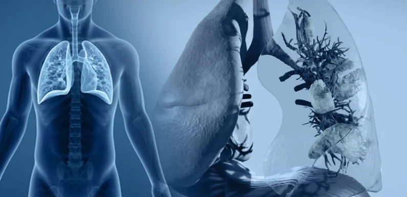
The lungs perform one of the body’s most essential and continuous tasks, exchanging oxygen and carbon dioxide with every breath. When that exchange breaks down, the consequences can escalate quickly, affecting nearly every organ system that depends on adequate oxygen delivery. Respiratory failure is among the most common reasons patients require intensive care, and understanding its types, causes, and the role of mechanical ventilation is essential knowledge for anyone involved in critical care.
What Respiratory Failure Means
Respiratory failure occurs when the respiratory system can no longer maintain adequate gas exchange to meet the body’s metabolic demands. This can mean insufficient oxygen reaching the bloodstream, an inability to remove carbon dioxide effectively, or both occurring simultaneously. It can develop suddenly, as in acute respiratory failure following an injury or infection, or gradually, as seen in chronic conditions like advanced chronic obstructive pulmonary disease, where the body has had time to partially compensate.
Type 1 Versus Type 2 Respiratory Failure
Respiratory failure is generally classified into two main types based on blood gas patterns. Type 1, or hypoxemic respiratory failure, is characterized primarily by low blood oxygen levels with normal or low carbon dioxide levels. It commonly results from conditions that impair oxygen exchange at the level of the alveoli, such as pneumonia, pulmonary edema, or acute respiratory distress syndrome. Type 2, or hypercapnic respiratory failure, involves elevated carbon dioxide levels, often alongside low oxygen, and typically results from inadequate ventilation, meaning the lungs are not effectively moving air in and out. This pattern is commonly seen in conditions that impair the mechanics of breathing, such as severe asthma exacerbations, neuromuscular disorders, or opioid-induced respiratory depression.
Common Causes of Acute Respiratory Failure
A wide range of conditions can precipitate acute respiratory failure. Pneumonia and other lower respiratory infections remain among the most common triggers, alongside acute respiratory distress syndrome, which can develop following sepsis, trauma, or severe infections. Pulmonary embolism, exacerbations of chronic lung disease, pulmonary edema related to heart failure, and severe asthma attacks are additional frequent causes. Neuromuscular conditions that weaken the muscles responsible for breathing, drug overdoses that suppress the respiratory drive, and chest trauma can all impair the body’s ability to ventilate effectively as well.
Recognizing the Signs of Respiratory Failure
Patients experiencing respiratory failure often present with shortness of breath, rapid or labored breathing, and use of accessory muscles to assist with each breath. Skin color changes, including a bluish tint around the lips or fingertips, indicate significant oxygen deprivation. Confusion, restlessness, or decreased level of consciousness can occur as oxygen delivery to the brain becomes compromised, while excessive sleepiness or lethargy may signal rising carbon dioxide levels. Rapid heart rate, abnormal breath sounds, and in severe cases, an inability to speak in full sentences due to breathlessness, are additional warning signs that warrant urgent evaluation.
Diagnosing Respiratory Failure
Diagnosis typically relies on arterial blood gas analysis, which directly measures oxygen and carbon dioxide levels along with blood pH, providing a clear picture of the type and severity of respiratory failure present. Pulse oximetry offers a quick, noninvasive estimate of oxygen saturation, though it does not capture carbon dioxide levels. Chest imaging, such as X-rays or CT scans, helps identify underlying causes like pneumonia, pulmonary edema, or pneumothorax, while laboratory tests and clinical history help narrow down the specific trigger requiring treatment.
The Role of Mechanical Ventilation
When a patient’s own respiratory effort cannot maintain adequate gas exchange, mechanical ventilation provides external support to take over or assist the work of breathing. Noninvasive ventilation, delivered through a tightly fitted mask, can be effective for certain patients, particularly those with chronic lung disease exacerbations or early-stage respiratory distress, and avoids the risks associated with more invasive methods. Invasive mechanical ventilation, requiring placement of an endotracheal tube connected to a ventilator, is reserved for more severe cases or situations where noninvasive methods are insufficient or contraindicated.
Ventilator settings can be adjusted across several modes, ranging from fully controlled breaths for patients unable to initiate their own respiratory effort to modes that support and supplement a patient’s spontaneous breathing. Clinicians must continually balance providing adequate oxygenation and ventilation against the risks of ventilator-associated complications, including lung injury from excessive pressure or volume, and prolonged immobility.
Weaning From Mechanical Ventilation
As a patient’s underlying condition improves, the goal shifts toward safely weaning them off ventilator support. This process typically involves gradually reducing ventilator assistance while closely monitoring the patient’s ability to breathe independently, along with assessing strength, mental status, and the resolution of the original cause of respiratory failure. Premature extubation can lead to respiratory distress and the need for reintubation, while unnecessarily prolonged ventilation increases the risk of complications, making this transition a carefully managed clinical decision.
Why Continued Clinical Education Matters
Given how quickly respiratory failure can progress and how many underlying causes can trigger it, clinicians benefit from regularly reinforcing their understanding of assessment, diagnosis, and ventilator management. A focused course on respiratory failure and mechanical ventilation can help solidify these concepts for practical application.
Final Thoughts
Respiratory failure is a complex, often rapidly evolving condition that requires prompt recognition and a clear understanding of its underlying cause to guide effective treatment. Whether managed with supplemental oxygen, noninvasive support, or full mechanical ventilation, the goal remains the same: restoring adequate gas exchange while giving the underlying condition time to resolve. Critical care nurses and respiratory clinicians can earn nursing CEUs in mechanical ventilation and respiratory assessment as part of their continuing education requirements, ensuring that professional development time is invested in the high-acuity knowledge most directly applicable to ICU and acute care practice.
